Maxwell E. Miller

Marine Biodiscovery

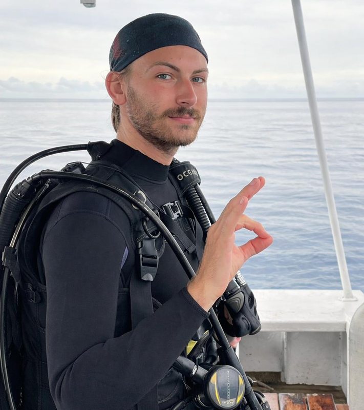
Portrait of Maxwell E. Miller in scuba equipment on the ocean

Marine scientist and biochemist interested in bioprospecting in the deep sea and extreme marine habitats. Joining University of Florida’s Whitney Laboratory for Marine Bioscience in August 2026 to investigate biomineralization and help develop the sea anemone Nematostella vectensis as a platform for protein expression.

Research directions

Incoming work at Whitney

Beginning August 2026

Beginning in August 2026, I will work with Drs. Mark Martindale and Sandra Loesgen at the Whitney Laboratory for Marine Bioscience. The project will investigate biomineralization pathways and help develop the sea anemone Nematostella vectensis as a platform for protein expression. The longer-term vision is to determine whether a genetically tractable anemone can reproduce aspects of coral-like biomineralization, informally, to “turn an anemone into a coral.” More broadly, I am interested in how marine organisms and their molecular machinery can become new research and biotechnology tools.

Working with Dr. Martindale will deepen my understanding of cnidarian development, evolution, and marine model systems. Dr. Loesgen’s expertise in natural products and organic chemistry will provide a complementary chemical perspective, creating a particularly strong environment for connecting organismal biology with marine biodiscovery.

Featured publication

Plain-language summary

Mucolipidosis IV is a rare inherited disorder that causes neurological impairment and progressive vision loss. This study tested a gene therapy designed to carry a functional copy of the affected gene across the blood-brain barrier in mice that were already showing symptoms. The treatment improved neurological function, reduced brain pathology, and prevented paralysis. Although the therapy reached the retina, it did not restore retinal thickness.

My contribution

I performed in vivo spectral-domain optical coherence tomography imaging of treated and control mice, segmented retinal layers, and analyzed retinal-thickness data to evaluate the therapy’s effects on retinal structure.

Methods spectral-domain OCT · retinal-layer segmentation · in vivo image acquisition · quantitative data analysis

Education & honors

Get in touch

If we cross paths at ASP 2026, or you work in natural products and biodiscovery, I would be glad to connect.